Basic Research Briefs
Reversing Lead Poisoning’s Effects
By Edyta Zielinska, Merrill Meadow | Illustrations by Jamie Cullen

It’s estimated that more than 500,000 U.S. children have so much lead in their blood that they are at risk of developing severe cognitive and behavioral impairments. But a major preclinical study offers hope that most of the genetic changes associated with those impairments could be reversed.
“Our new research shows that lead poisoning in children affects a wide array of genes in the part of the brain involved in learning and memory,” explains neuroscientist Jay Schneider, PhD. “However, our data suggest that enriched early childhood environments and access to stimulating activities could minimize or potentially reverse those effects.”
Dr. Schneider’s team studied rats exposed to lead in a way that replicated conditions and developmental timing of children exposed to lead in early childhood. After the lead exposure, the rats were separated into two different housing conditions: “enriched” cages, with substantial opportunities for interaction with other rats and with a variety of toys, climbing and nesting materials, and tunnels that were changed regularly; and cages offering much less stimulation and social interaction. Then the team assessed gene expression in the brains of both groups.
The researchers found that lead exposure affected the expression of more than 3,500 genes. As much as 80% of these lead-induced gene expression changes were reversed in rats in the enriched or high-stimulation environment.
The researchers agree that their study clearly points to the neurological benefit of providing at-risk children and children known to be exposed to lead with stimulating environments and rich social interactions.
Eye-Disease Defenses
By Karuna Meda, Merrill Meadow | Illustrations by Jamie Cullen

Our eyes need to protect themselves from many things — ranging from dust and chemicals to bacteria and ultraviolet light. Yet scientists long believed that key parts of the eye lack protection by immune cells.
Then, a few years ago, cell biologist Sue Menko, PhD, and collaborating researchers observed immune cells trying to fix the damaged lens of mice they were studying. They found that when mouse corneas were wounded, immune cells travelled to the surface of the lens to repair the eye and protect from further damage.
The researchers wondered whether they would see the same kind of responses in uveitis, an auto-immune inflammatory disease of the eye. Even though, in humans, uveitis can lead to retinal scarring, glaucoma and cataracts of the lens, the role of immune cells in uveitis-associated cataracts had never been explored. “That’s why, when our team used high-resolution microscopy to observe the uveitis-model mice, what we saw amazed us: a robust response comprising dozens of immune cells of different types, including T-cells and macrophages,” says Dr. Menko.
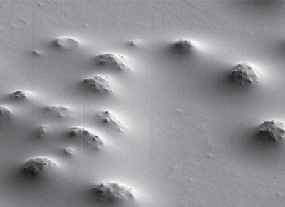 The researchers used a scanning electron microscope to capture detailed changes on the surface of the lens capsule of the eye. They found many bumps, indicating regions where immune cells had become integrated within the lens capsule in response to damage and inflammation. (Image credit: Mary Ann Stepp, PhD, George Washington University.)
Moreover, they found that the lens capsule — a thick protein-rich layer surrounding the lens — had deep pits or divots. “These formations are evidence that the immune cells are actually integrating into the lens capsule,” explains JodiRae DeDreu, a PhD student researcher in the Menko lab. In fact, they observed the number of divots increasing and becoming deeper, indicating further invasion of immune cells into the lens capsule as the disease progressed.
“There is much work still to be done,” says Dr. Menko. “But we believe that uncovering these processes — which had never been shown before — could lead to wholly new pathways for understanding and treating eye disease.”