Wu Research
Contact
909 Walnut Street
1st Floor
Philadelphia, PA 19107
Dr. Chengyuan Wu is a board-certified fellowship-trained Stereotactic and Functional Neurosurgeon with a background in the fields of Computer and Biomedical engineering. For this reason, he is uniquely positioned in the field of neuroscience research. Specifically, he is able to blend his knowledge of signal and image processing techniques with his clinical expertise in Neurological Surgery.
His work began during his graduate education at Tufts University School of Engineering, where he focused his thesis on the subject of signal processing for automated analysis of electroencephalograms (EEGs). For more than a decade, he has continued to dedicate his research efforts to the fields of epilepsy and movement disorders.
As part of the Comprehensive Epilepsy Center at Thomas Jefferson University, Dr. Wu has had an opportunity to be part of a clinically busy practice, which performs approximately 100 surgeries for epilepsy each year covering the spectrum from traditional resective surgeries and invasive implantations to minimally invasive interventions and neurostimulation. He routinely integrates advanced neuroimaging and image post-processing to deliver the highest level of care to his patients.
As part of the Comprehensive Parkinson’s Disease & Movement Disorder Center at Thomas Jefferson University, Dr. Wu and his team perform approximately 60 deep brain stimulation (DBS) implants each year. Dr. Wu also applies advanced imaging techniques to patients undergoing DBS for movement disorders such as Parkinson's Disease and Essential Tremor.
Dr. Wu continues to work on translational research focused on advancing the field of neuroimaging in order to not only improve the future of epilepsy and DBS surgery, but also to further our understanding of the brain and the networks embedded within.
Research Projects
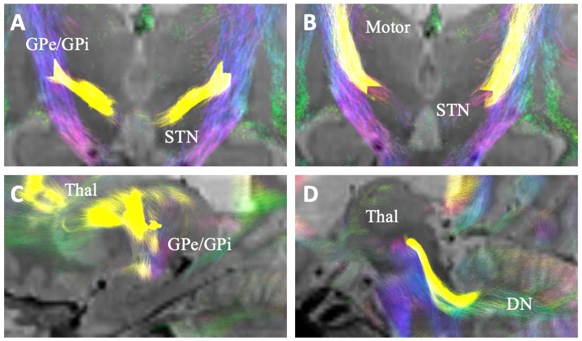
Investigation of Tractography Based Programming for Deep Brain Stimulation
An area in which Dr. Wu sees immense potential for development in his specialty is in the ability to discover how tailoring treatments to each one of his patients can improve their care. This hypothesis has led him to seek a method of individualized treatment when treating Parkinson’s Disease through Deep Brain Stimulation. In collaboration with a skilled team at Duke University led by Dr. Cameron McIntyre, our research investigates the efficacy of DBS in patients affected by Parkinson’s Disease through implementation of pathway-based programming in comparison with traditional, standardized DBS programming. Each tractography based program is created using the neuroanatomy of the patient being treated, MRI information, and DBS lead location. This study intends to both elucidate the specifics of the workings of brain connectomes as well as provide knowledge to allow for innovative clinical treatments to make DBS more effective and efficient for each patient.
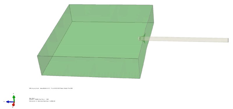
Modeling of Brain Deformation in Deep Brain Stimulation Surgery with a Combination of Magnetic Resonance Imaging, Force/Torque Signals, and Numerical Simulation Methods
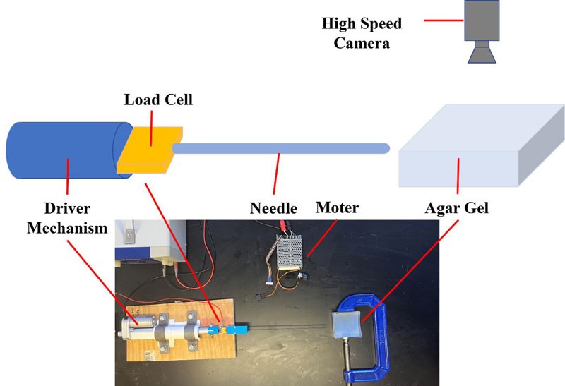
Together with biomedical researchers at Villanova University, our lab is working to create a model that will allow us to predict how an individual’s brain will shift and react to neurosurgical procedures, specifically Deep Brain Stimulation. Being able to anticipate deviations within brain matter that will occur during surgery has the potential to greatly improve accuracy down to the millimeter and allow for safer and more effective procedures. By incorporating force measurements and MRI imaging techniques such as MR elastography (MRE), a technology which allows for measurement of tissue stiffness through low-frequency vibrations, we plan to create a simulation that allows us to gain a deeper understanding of how the brain may move in response to different stimuli.
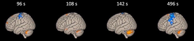
Investigation of Deep Brain Stimulation on Connectome Utilizing Functional MRI Imaging
Deep Brain Stimulation (DBS) is an incredibly effective treatment for diseases and disorders such as Parkinson’s Disease and Essential Tremor, yet there is still much to be discovered about the intricate ways that it affects the brain and provides relief to patients. Blood Oxygen Level Dependent (BOLD) imaging forms the basis of functional magnetic resonance imaging (fMRI), which has great potential to identify distributed associative neural networks and reveal features of brain organization. By comparing fMRI scans of patients with implanted DBS systems while their systems are turned on and turned off, our lab hopes to discover more about how DBS influences connections within the brain and what that may reveal about functional networks that allow for such profound improvement in the quality of life for patients suffering from Parkinson’s Disease, Essential Tremor, and other movement disorders.