
Mantegazza Research
Jefferson Alumni Hall, (Office)
Jefferson Alumni Hall, (Lab)
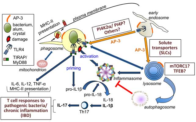
The Mantegazza lab studies pro-inflammatory pathways in Dendritic cells (DCs) – the main antigen-presenting cells and instructors of T cell responses, with a focus on phagocytosis (the capture of bacteria or particles) and lysosomal function. We are interested in investigating the cross-talk between phagosomes and inflammasomes and identifying new regulators of phagosome maturation, inflammasome activity and autophagy with the ultimate goal of tuning immune responses in DCs. After phagocytosis, phagosomes mature through a series of interactions within the endo-lysosomal system. This process, known as phagosome maturation, promotes, via Toll-like receptor (TLR) signaling, other cellular processes, such as antigen presentation, proinflammatory cytokine production and inflammasome priming, all key players in immune responses. Inflammasome activity – the “big bang” of inflammation triggered by pathogens or self-damage –, is regulated by phagosomal TLR signaling and autophagy. Autophagy, in turn, may dampen inflammasome activity, keeping inflammation under control. Previously, we had identified adaptor protein AP-3 as a regulator of phagosomal TLR4 signaling, inflammasome activity and autophagy. However, the role of AP-3 is indirect. We are investigating more direct regulators of these pathways – such as the family of phago-lysosomal solute transporters, SLCs –, with the aim of tuning immune responses in DCs, to ensure proper inflammatory responses to pathogens and prevent chronic inflammation that may lead to disease (i.e. inflammatory bowel disease).
Research Projects
Role of phosphoinositide platforms in promoting TIRAP/MyD88- dependent TLR signaling from phagosomes
The coordinated activity of lipid kinases and phosphatases is required for the formation of lipid platforms conducive to the recruitment of TLR sorting adaptors such as TIRAP to the phagosome, to promote TLR signaling. Phagosomal TLR signaling is essential for the formation of phagosomal tubules, antigen MHC-II presentation and proinflammatory cytokine secretion in DCs, to ensure efficient anti-microbial responses. One of the regulators of phagosomal TLR4 signaling is the lipid kinase PI4K2a, and its lipid, PI4P, by ensuring TIRAP docking and supporting MyD88-dependent TLR signaling. We are investigating additional regulators required for optimal phagosomal TLR4 signaling and downstream anti-microbial immune responses, using a combination of genetic and biochemical approaches, live-cell imaging and immunoassays. Our ultimate goal is to identify targets amenable to modulation to optimize the anti-microbial response in DCs.
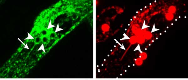 Bone marrow-derived DCs transduced with PI4K2a-GFP and pulsed with OVA-Txred beads. Arrowheads indicate phagosomes; arrows indicate phagotubules. Phagotubules are induced by phagosomal TLR signaling and promote phagosome cross-talk and MHC-II presentation in DCs. PNAS 2020 Nov. 10; 117(45).
Role of phagolysosomal solute carrier transporters (SLCs) in inflammasome activity and autophagy
Solute transporters (SLCs) present on phagolysosomes sense nutrients and phagosome degradation products and convey nutrient sufficiency to the lysosomal complex mTORC1, the master regulator of autophagy and cell growth. SLC function is associated with human inflammatory disorders such as Lupus and inflammatory bowel disease (IBD), highlighting the importance of phagosomal nutrient sensing/ signaling in the regulation of immune responses. We are investigating how mTORC1 signaling impacts inflammasome activity after infectious or sterile stimuli by regulating inflammasome positioning and inactivation by autophagy, using a combination of immunofluorescence microscopy (confocal and spinning-disk), biochemical and flow cytometric approaches and immunoassays. We are also investigating the role of SLCs in preventing chronic inflammation by the modulation of inflammasome activity and autophagy in a mouse model of IBD. The understanding of the molecular mechanisms that regulate inflammasome activity will allow the tuning of immune responses, by boosting immune responses to pathogens and dampening excessive inflammation.
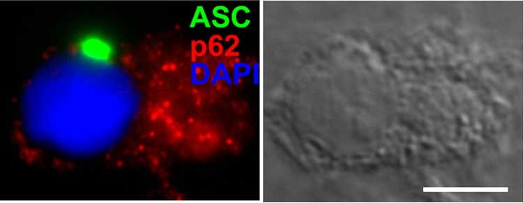 Inflammasome.jpg. Figure legend: Bone marrow-derived DCs transduced with ASC-GFP (inflammasome component), infected with Salmonella Typhimurium and stained with anti-p62. The activation of the inflammasome results in the convergence of ASC-GFP fluorescence in a single spot known as the inflammasome “speck”. p62 is used as a marker for autophagy induction. Note that the speck appears to be protected from autophagy. PLoS Pathogens 2017 Dec. 18; 13(2).
How lipid-based vaccine adjuvants activate Dendritic Cells
Vaccine adjuvants enhance and prolong immune responses elicited by vaccine antigens. In collaboration with the Schnell and Kurup Labs., we are investigating how certain vaccine adjuvants activate pro-inflammatory signaling, inflammasome activity and autophagy, and modulate antigen presentation in DCs.