Stroke remains the leading cause of disability and the fifth leading cause of death in the US. It is now widely accepted that following stroke, endogenous neural stem cells (NSCs) residing in the classic stem cell niches of the adult brain, the SVZ and SGZ, differentiate into new neurons which may be important for repair of the rat and human brain. We recently found that stem cell niches in midline circumventricular organs (CVOs) and at other sites along the cerebral ventricles, where stem cells are greatly amplified, leading to enhanced neurogenesis, after stroke. The same regions also possess a highly permeable blood-brain-barrier (BBB). We hypothesized that access to factors in blood after stroke can drive these events in the stem cell niche. Our goals are: 1) defining the molecular pathways used by those systemic blood factors to regulate stem cell activities; 2) studying functional changes in BBB permeability in stem cell niches at various time intervals after stroke; 3) studying whether elevated VEGF induces stem cell proliferation and differentiation in brain niches after stroke.

Iacovitti Research
Contact
900 Walnut St
461 JHN
Philadelphia, PA 19107
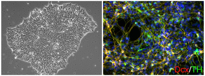
Our lab focus is in the fields of stem cell biology and neuroprotection with an emphasis on Parkinson’s disease (PD) and stroke models. Human induced pluripotent stem cells (iPSCs, left), with their potential to generate autologous patient-derived cells (such as, dopamine neurons in green, right) hold great promise for the study and treatment of a host of devastating diseases, including PD. Endogenous stem cells present in the adult brain are likewise important in repair particularly after acute injury like stroke. Our lab has concentrated on the molecular and cellular changes in the blood-brain-barrier that ultimately lead to regulation of stem cell proliferation and neuronal differentiation after stroke. We also have focused our studies on the discovery of novel factors capable of protecting neurons at risk for dying during progressive disease (PD) or after injury (stroke).
Research Projects
Differentiating stem cells into dopaminergic neurons to study and treat Parkinson's Disease
Our lab studies focus on the differentiation of CNS neurons from stem cells, in particular, the dopamine neuron. These studies include epigenetic and environmental regulation of cell phenotype. Our goals are 1) defining the underlying principles governing the differentiation of the midbrain dopamine neuron, the cell lost in Parkinson’s disease (PD); 2) using differentiated patient- specific iPS cells for PD modeling and drug discovery in culture; and 3) translating these discoveries into therapies to treat PD in patients.
Protecting Dopamine Neurons from PD toxins with Astrocytes
PD is a well-studied neurodegenerative disease which is characterized by the selective degeneration of dopamine neurons of the substantia nigra pars compacta (SN), while the DA neurons of the neighboring ventral tegmental area (VTA) are relatively spared. In addition to studying intrinsic differences between SN and VTA neurons, our lab also examines neighboring astrocytes, which are known to release a variety of neurotrophic factors. We recently showed that the astrocytes from these two regions are regionally specified, with VTA astrocytes uniquely producing the growth factor GDF-15 which can rescue both SN and VTA DA neurons from PD challenge. These findings raise the intriguing possibility that the vulnerability of specific populations of neurons to disease risk factors may not stem entirely from neuron-intrinsic susceptibilities but also from differences in support from their local astrocytic microenvironments.
Are microglia regionally specified and how do they contribute to PD vulnerability?
In addition to midbrain neurons and astrocytes, microglia (MG), the resident immune cells of the brain, likely play a key role in the degeneration of SN neurons in PD. Intriguingly, SN houses greater numbers of MG and fewer astrocytes while the neighboring VTA is characterized by low numbers of MG and a high number of astrocytes. Is life or death then a balancing act between damaging MG and protective astrocytes? Using PD iPSC models and the hTH-GFP reporter rat we would like to answer: 1) Are MG regionally distinct like midbrain astrocytes and neurons, expressing specific cell surface markers, transcription factors, cytokines, etc.? 2) Can we adapt what we learn from the rat model to a human iPSCs system? 3) Can we examine the roles of neurons, astrocytes, MGs in PD susceptibility using PD patient-specific iPS cells?
Stroke and its effects on brain stem cells and stem cell niches
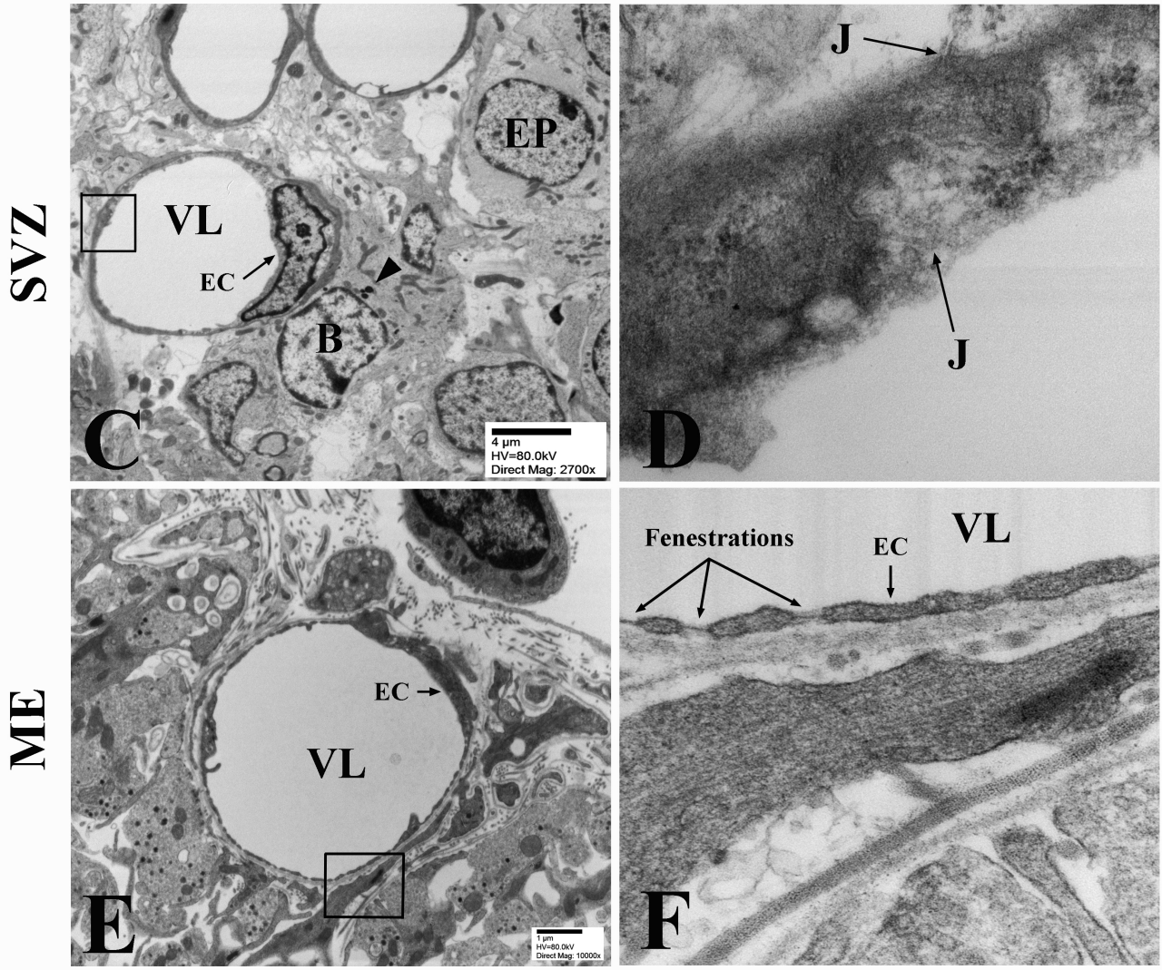 Disrupted tight junction after stroke leads to BBB permeability change in stem cell niches.
Opening the BBB to potential therapeutics
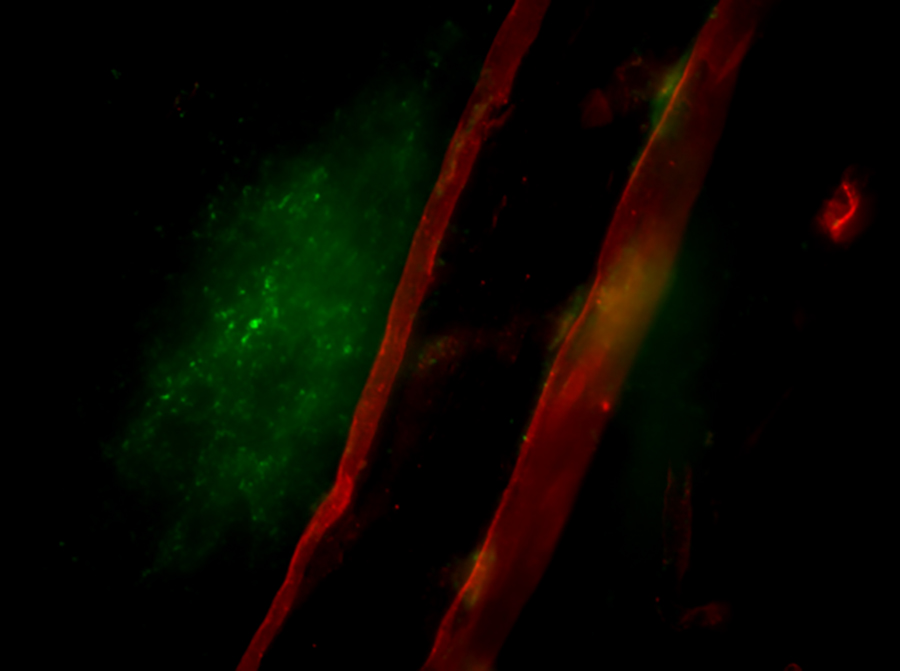 Extravasation of 70kDa FITC-labeled Dextran out of cerebral vasculature following rat sphenopalatine ganglion stimulation.
The blood-brain barrier (BBB) is a nearly impermeable wall between blood vessels and the brain. Composed of endothelial cells linked together by tight junctions, it is responsible for the accurate and efficient functioning of our brain. However, when it comes to treating pathological states, it is equally proficient at blocking the passage of therapeutic agents into the brain. We recently found that by stimulating the parasympathetic ganglion, called the sphenopalatine (SPG), the BBB can be transiently and reversibly opened for 20 minutes. Using this technique, we are able to move large molecules (70kD dextrans) and even bone marrow stem cells across the BBB into the brain. The potential impact of this work is far-reaching and game-changing for many diseases, including stroke, allowing us to get therapeutic drugs, antibodies, gene therapies and cell therapies into the brain. Currently we are studying the mechanism of opening the BBB.