Breast cancer preferentially metastasizes or migrates to the bones, where the five-year relative survival rate is less than 10%. Metastatic lesions are thought to originate from disseminated tumor cells (DTCs) shed from a primary tumor. Clinical evidence suggests that, upon entry in a secondary niche, DTCs undergo proliferative quiescence for a period of up to 25 years, where they are capable of escaping immune surveillance and evading chemotherapeutic agents which target cycling cells. As a result, breast cancer patients successfully treated for primary breast cancer decades before suddenly develop overt macrometastases without prior symptoms. This latency period before relapse at a secondary site can be defined as metastasis dormancy. Thus, there is a critical need for elucidating the molecular mechanisms facilitating breast cancer dormancy and reactivation in the bone such that preventative therapeutics can be developed. Despite the high prevalence of dormant tumors in humans, this area of cancer research is poorly understood and understudied. The Bussard laboratory studies how specific interactions between normal bone osteoblasts and breast cancer cells facilitate and maintain breast cancer cell dormancy in bone.

Bussard Research
Contact
233 South 10th Street
Bluemle Life Sciences Building, Room 624A
Philadelphia, PA 19107

Research Projects
Research Focus: The focus of research in the Bussard Laboratory is two-fold:
- Identify microRNA, proteins, and novel mediators that facilitate breast cancer cell dissemination to bone, including mediators of cancer cell arrival and localization, as well as delineate conditions that orchestrate proliferative quiescence and cancer cell re-activation in bone.
- In parallel elucidate the molecular basis of crosstalk between tumor cells and the host stroma, with a focus on understanding alterations in bone osteoblasts, that create a permissible environment for metastatic cancer cell colonization and survival. Research in Dr. Bussard’s laboratory utilizes both 2D and 3D in-vitro cell culture models, high-end fluorescence microscopy, novel humanized mouse models of tumor development, and implantable hydrogel biomaterial scaffolds to examine interactions between cancer cells and the host stroma, with a focus on bone metastatic breast cancer. Dr. Bussard is also the recipient of a NIH/NCI K99/R00 Pathway to Independence Award, which has been instrumental to establishing her research program.
Identification of Novel Mediators that Orchestrate Cancer Cell Dormancy in Bone.
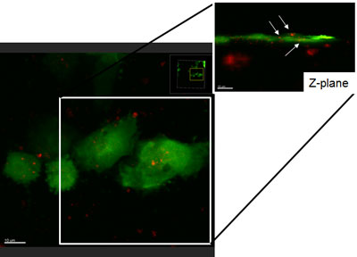
The goal of the first project in the Bussard Lab, supported by a R00, is to identify novel mediators which orchestrate disseminated breast cancer cell dormancy in the bone microenvironment (Figure 1). As a way to test this, we examine the roles of gap junctions and exchange of microRNAs between osteoblasts and disseminated breast cancer cells as modulators to this process. We have used three different models: cell culture, long-term three-dimensional culture, and animal models to study these interactions. We have found that osteoblasts form gap junctions with metastasis-suppressed breast cancer cells, which may contribute to cellular crosstalk between the two cell types. Bone osteoblasts were also found to produce exosomes which contain exosomal microRNAs found to be involved in cell cycle control and cellular dormancy. These osteoblast-derived exosomes were taken up by breast cancer cells in a co-culture setting.
Molecular Characterization of Tumor-Stromal Interactions in Metastasis.
The goal of a separate, independent project is to elucidate crucial tumor-stromal interactions between osteoblasts and breast cancer cells that drive metastatic progression. Using in-vitro and in-vivo models, we have found that breast cancer cells cause osteoblasts to alter their production of matrix proteins, and increase their production of a set of inflammatory cytokines: IL-6, IL-8, MCP-1, and VEGF. In fact, preliminary data have shown that, when treated with breast cancer conditioned medium, osteoblasts reduce their expression of normal bone turnover markers such as alkaline phosphatase, CD31, and alpha smooth muscle actin, yet increase their expression of matrix remodeling markers, including MMP13. These results are consistent with the described aggressive “matrix-remodeling” tumor-associated fibroblast. On the other hand, metastasis-suppressed breast cancer conditioned medium treated osteoblasts increased their expression of IL-6, suggestive of an osteoblast inflammatory response to disseminated breast cancer cells. Tumor-associated osteoblast cells also exhibited altered morphology in the form of a thin, spindle-like shape, as opposed to cobblestone appearance. These findings suggest that breast cancer cells educate osteoblasts into a tumor-associated fibroblast-like cell during the metastatic process.
Combined, these data suggest that there is extensive crosstalk between osteoblasts and disseminated cancer cells in bone, and highlights the importance of investigating osteoblasts as key players which mediate cancer cell proliferative quiescence in bone.