


Alizadeh-Mouchtouris-Research

Principle Investigators
- Lab Director
- Assistant Professor of Neurosurgery & Radiology
Jefferson Hospital for Neuroscience
900 Walnut Street, 2nd Floor, Room 226
Philadelphia, PA 19107

- Lab Clinical Co-Director
- Assistant Professor of Neurological Surgery
Jefferson Abington Hospital
1 Widener, 1200 Old York Road
Abington, PA
Precision Neuroimaging in Neurosurgery
Precision Neuroimaging in Neurosurgery (PNN), led by Mahdi Alizadeh, PhD, and Nikolaos Mouchtouris, MD, develops and evaluates imaging and electrophysiological methods for neurosurgical planning and the study of neurological disease. Our research combines diffusion and functional MRI with perfusion imaging, magnetic resonance elastography, and stereo-electroencephalography. Applications include spinal cord injury and neuropathic pain, chronic low back pain, and cervical myelopathy; vascular and tissue injury after stroke; neuromodulation for movement disorders, back pain, and epilepsy; and localization of epileptogenic networks. In collaboration with Jefferson’s neurosurgery, neurology, radiology, physical therapy, Wills Eye, and Moss-Magee Rehabilitation teams, we investigate how these measurements relate to anatomy, symptoms, and treatment outcomes.
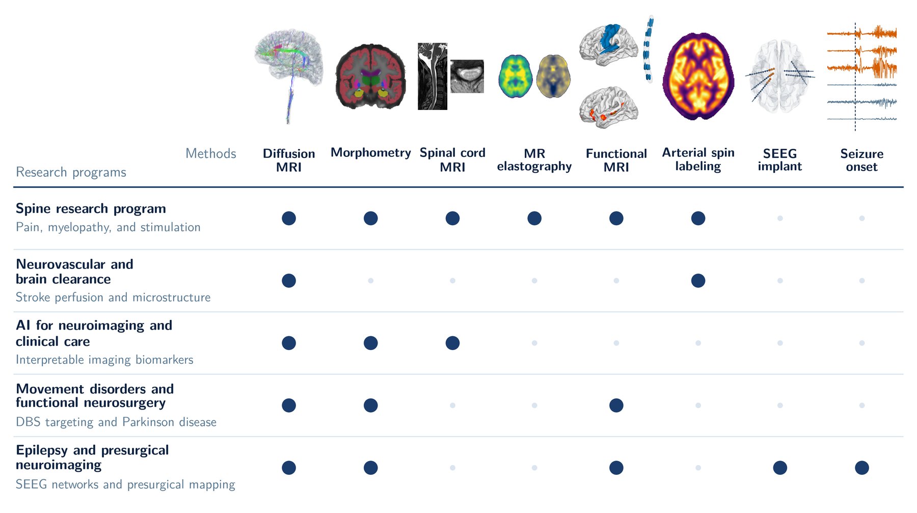 The lab’s methods (top) and the research programs that use them (dots). Tractography is from a healthy control of our R01 spinal cord injury pain study, shown in the brain and cervical cord. Other thumbnails use public data: FreeSurfer fsaverage (Fischl et al., 1999), spine-generic (Cohen-Adad et al., 2021), MRE134 (Hiscox et al., 2020), Yeo networks (Yeo et al., 2011), Individual Brain Charting (Pinho et al., 2018), ExploreASL (Mutsaerts et al., 2020), and OpenNeuro ds004100.
Click on images for larger version.
Research Projects
Spine Research Program
Spinal cord research is a major focus of the lab. We develop multimodal neuroimaging and quantitative PET-MRI to investigate the neural mechanisms of neuropathic pain after spinal cord injury supported by NIH/NINDS R01NS134987 as well as in those with cervical myelopathy and low back pain. We develop and validate quantitative MRI biomarkers for spinal cord injury and other spinal disorders, using reduced-field-of-view diffusion MRI, diffusion tensor imaging (DTI), spinal cord tractography, simultaneous brain and spinal cord resting-state fMRI, and structural and functional connectomics of brain-spinal cord.
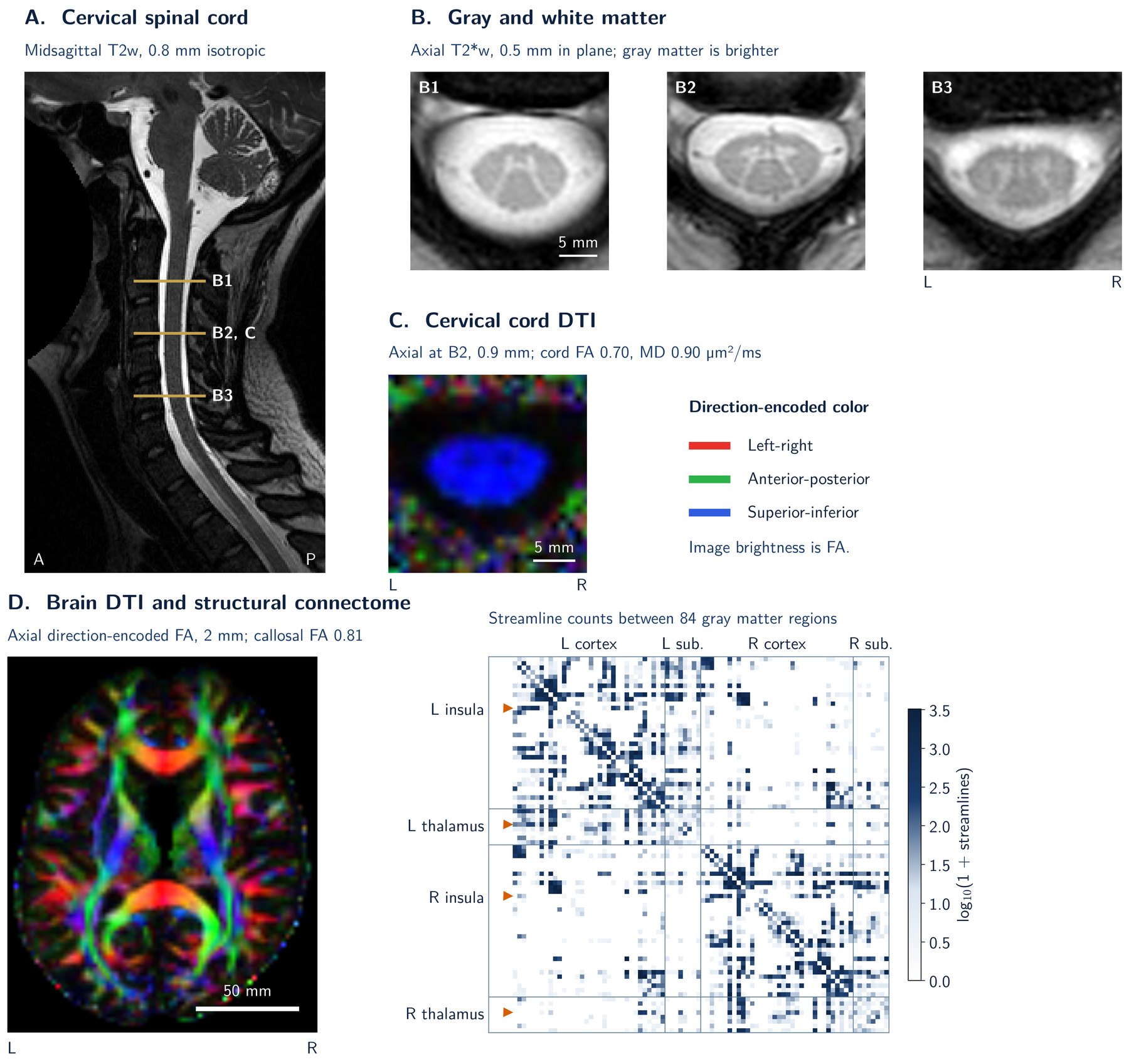 Brain and cervical spinal cord MRI. (A) Midsagittal T2-weighted image of the cervical cord, with the levels of the axial slices in B and C marked. (B) Axial T2*-weighted slices in which the gray matter butterfly is brighter than the surrounding white matter. (C) Cord diffusion tensor imaging (DTI) at level B2 in direction-encoded color; brightness is fractional anisotropy (FA). (D) Brain DTI and a structural connectome of streamline counts between 84 gray matter regions; the insula and thalamus, regions implicated in neuropathic pain, are marked. A-C are from one healthy participant of the spine-generic dataset (Cohen-Adad et al., 2021; CC BY 4.0), and D is from the Stanford HARDI dataset (Rokem et al., 2013; PDDL 1.0).
Neurovascular & Brain Clearance Research
This program combines Dr. Mouchtouris’s cerebrovascular and endovascular research with Dr. Alizadeh’s expertise in quantitative MRI. We study how the vascular system, brain physiology, and tissue microstructure relate to neurological dysfunction, using quantitative perfusion MRI, imaging of the blood-brain and blood-spinal cord barriers, and diffusion-based evaluation of perivascular spaces and brain clearance. We integrate these noncontrast measures with structural and functional MRI and develop them as biomarkers of vascular and tissue physiology. Current work focuses on ischemic stroke and its treatment and functional outcome.
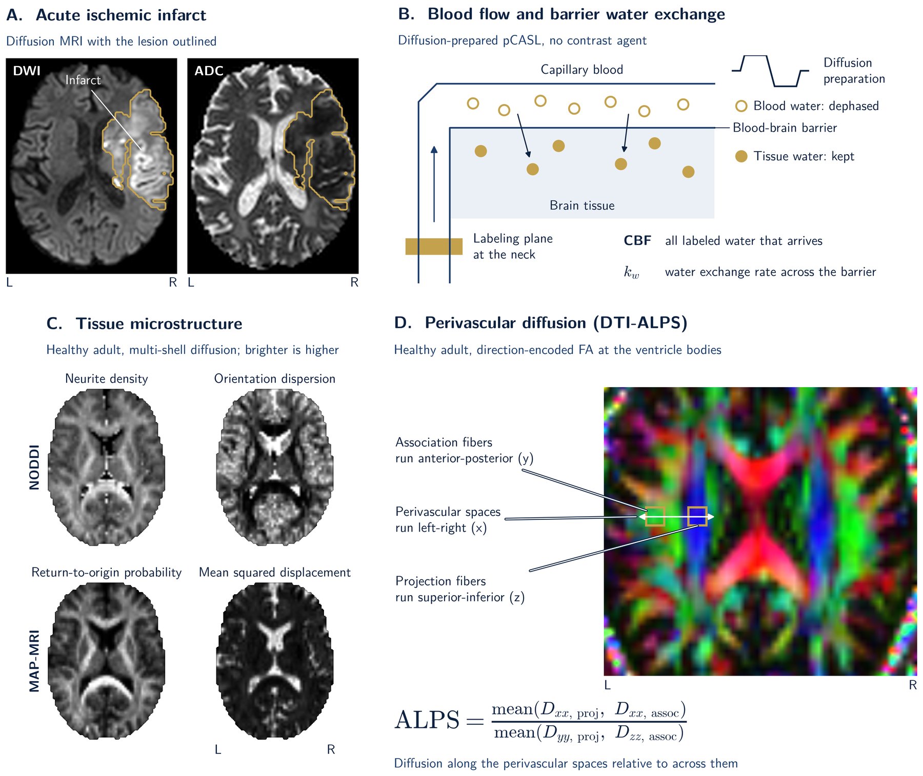 Noncontrast imaging of vascular and tissue injury after acute ischemic stroke. (A) An acute infarct on diffusion-weighted imaging (DWI) and the apparent diffusion coefficient (ADC) map, with the lesion outlined. (B) Diffusion-prepared pseudo-continuous arterial spin labeling (DP-pCASL) measures cerebral blood flow (CBF) and water exchange across the blood-brain barrier (kw) without a contrast agent. (C) Neurite orientation dispersion and density imaging (NODDI) and mean apparent propagator (MAP) MRI describe tissue microstructure. (D) The index of diffusion tensor imaging analysis along the perivascular space (DTI-ALPS) compares diffusion along the perivascular spaces with diffusion across them, beside the bodies of the lateral ventricles. We relate these measures to thrombectomy and to disability on the modified Rankin Scale. Images are illustrative, from public datasets: ISLES 2022 (Hernandez Petzsche et al., 2022; CC BY 4.0) in A and a healthy adult multi-shell dataset (Hansen and Jespersen, 2016; CC0) in C and D.
AI for Neuroimaging & Clinical Care
Our machine learning research spans the development of new artificial intelligence (AI) methods and their translation into neurosurgical practice. We design deep learning models that extract diagnostic and prognostic information from medical images and infer measurements that would otherwise require specialized acquisitions. In partnership with Jefferson clinical teams, we also build AI systems that support clinical decision-making and care delivery.
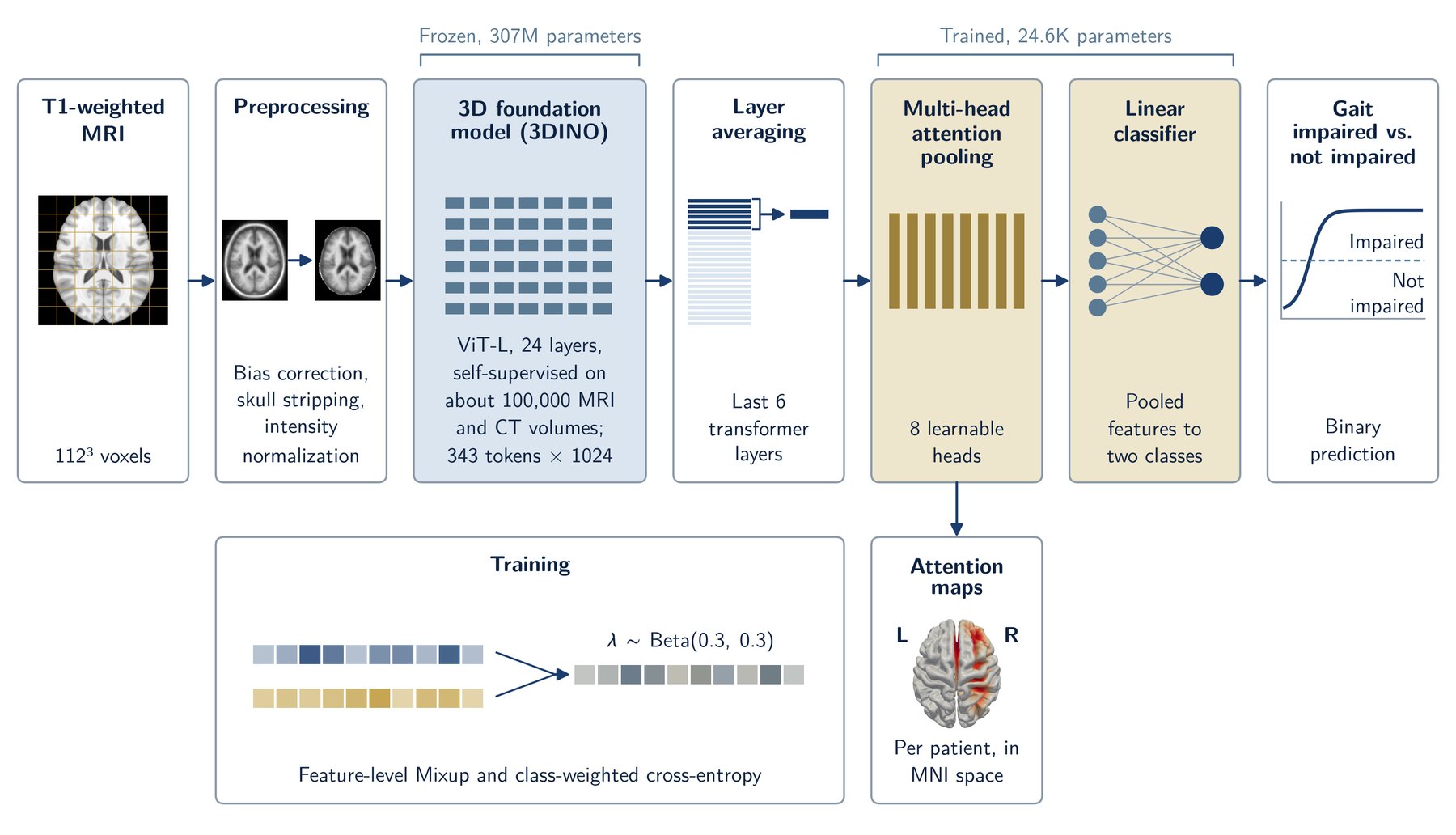 Architecture of the interpretable vision transformer for classifying gait impairment in Parkinson’s disease from a single T1-weighted MRI. Patch-token features from a frozen, self-supervised 3D foundation model (3DINO; Xu et al., 2025) are averaged over the last six transformer layers and summarized by multi-head attention pooling, and a linear classifier predicts gait impairment. Only the pooling and classification layers are trained. The attention weights yield patient-specific maps that are registered to MNI space. The surface shows the mean attention map of correctly classified gait-impaired patients in a held-out test set, projected onto the FreeSurfer fsaverage template (Fischl et al., 1999) in dorsal view; the input slice is from the MNI152 template (Fonov et al., 2011).
Movement Disorders & Functional Neurosurgery
We investigate imaging methods for deep brain stimulation (DBS), including target definition, lead localization, and factors associated with electrode deviation. Related studies examine the structural and functional abnormalities associated with gait and visual symptoms in Parkinson’s disease.
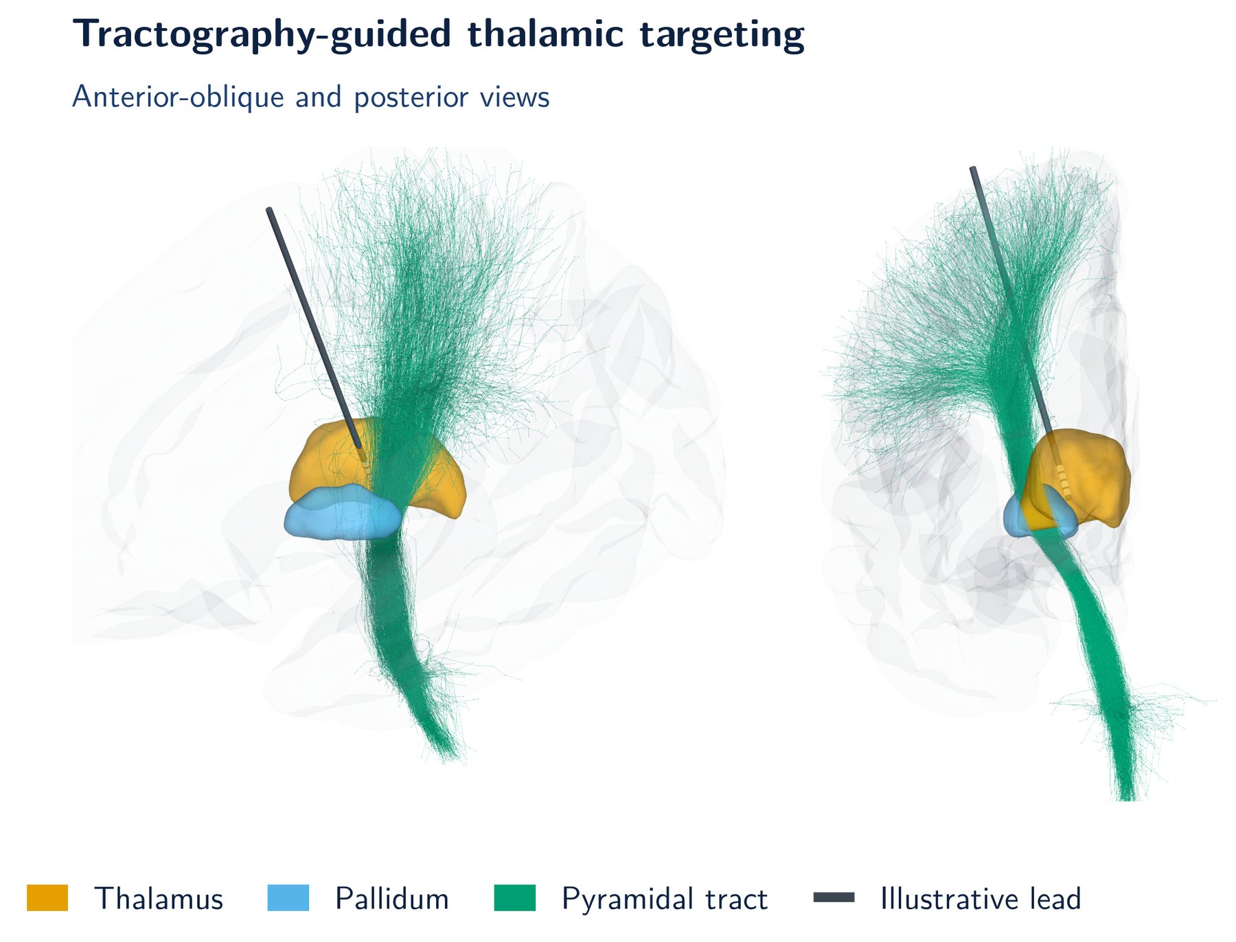 Tractography-guided thalamic targeting for deep brain stimulation, shown in anterior-oblique (left) and posterior (right) views. A left pyramidal-tract reference bundle (Rheault, 2019; CC BY 4.0) is displayed with the left thalamus and pallidum from the FreeSurfer fsaverage template (Fischl et al., 2002). The lead, with four ring contacts in the ventral thalamus, is an illustrative drawing, not a planned or implanted trajectory.
Epilepsy & Presurgical Neuroimaging
We integrate stereo-EEG, structural MRI, diffusion imaging, and functional MRI to investigate epileptogenic networks in drug-resistant epilepsy and support surgical planning. Work examines interictal electrophysiological markers, neural fragility, temporal network features across wakefulness and sleep, and associations with temporal lobectomy outcomes. A preliminary 22-patient study related the spatial distribution of beta/high-gamma correlations to postoperative seizure freedom. These methods are being investigated as candidate markers for seizure localization and outcome assessment.
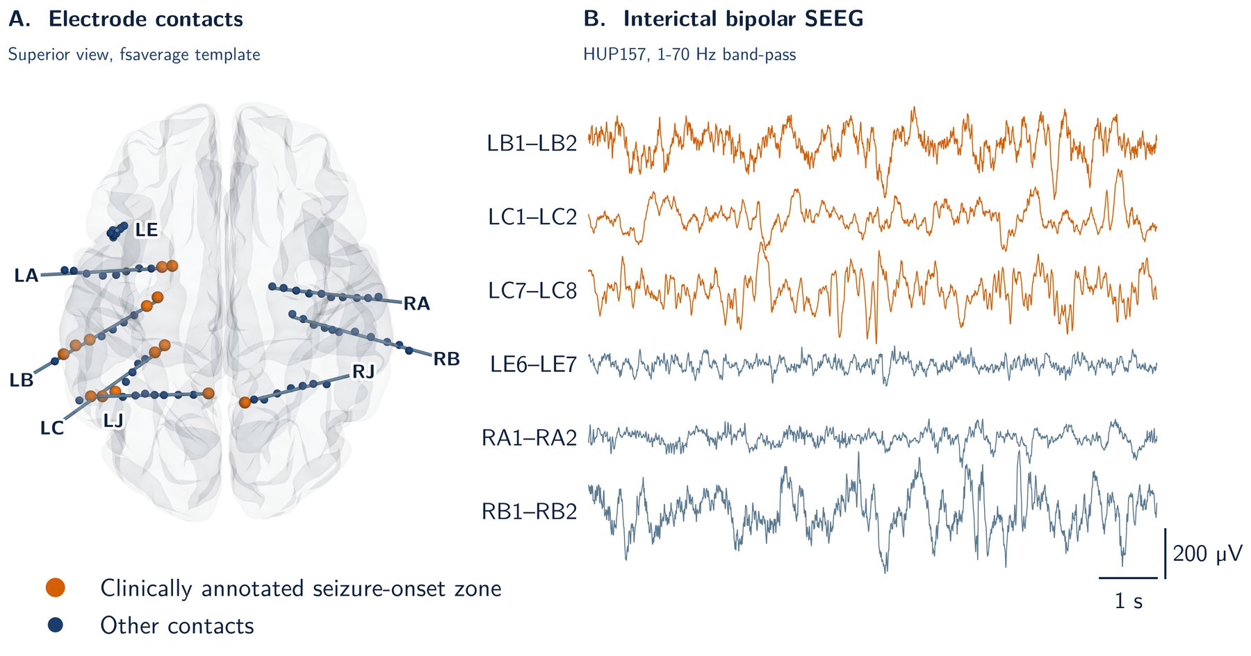 Stereo-EEG in drug-resistant mesial temporal lobe epilepsy (patient HUP157, OpenNeuro ds004100, CC0). (A) Depth-electrode contacts normalized to the fsaverage template, superior view with anterior at the top and left on the left; contacts in the clinically annotated seizure-onset zone are highlighted, and shafts are drawn as straight lines fitted to the contacts. (B) Ten seconds of interictal bipolar recordings from selected contacts, band-pass filtered at 1-70 Hz.