")
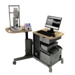
Optical Coherence Tomography (OCT) is a non-invasive, non-contact imaging modality used to visualize and monitor changes to the morphology of biological tissue. OCT employs low-coherence interferometry to create cross-sectional images that reveal sub-surface details of the tissues of interest. In the most common ophthalmic applications OCT systems use near-infrared light to generate high-resolution, volumetric images of tissue microstructures including the cornea, iris, crystalline lens, vitreous and retina.
The Department possesses both the Leica Bioptigen and Heidelberg Spectralis instruments to meet your imaging requirements.