
Atomic Force Microscopy
Mission, Goals, Capabilities
Atomic force microscopy (AFM) utilizes a rigid cantilever to probe the surface of samples with nanoscale resolution – far surpassing the optical diffraction limit.
Users typically utilize AFM for one of three major applications:
- Quantification of tissue nanomechanical properties
e.g. tissue stiffness) - Determination of topographical information
(e.g. surface roughness), or ... - Quantification of nanoscale interactions
(e.g. attaching an antibody to the AFM probe)
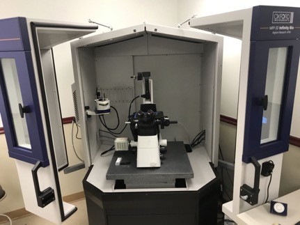
Our AFM system also includes an inverted fluorescence microscope, which can be utilized simultaneously to locate structures or cells that have been fluorescently tagged. In addition, our system can perform “Fast Force Mapping”, which is technique to rapidly capture a force-displacement curve at every pixel in the field of view. With an extended Z-axis, surface features up to 40 microns can be probed. Samples may be stiff (e.g. bone, metal, plastic) or compliant (e.g. cells), but must be physically attached to either a slide or a cell culture dish. However, imaging can be performed in either air or liquid. For lengthy in vitro experimentation, our system is equipped to provide temperature control for cell culture.
Major Equipment
Reserve equipment through iLab.
- Oxford Instruments MFP-3D Infinity BIO integrated with a Nikon inverted fluorescence microscope
Services
- Nanomechanical properties
- Surface roughness and topology
- Custom applications