Solving the Puzzle of Fibrotic Diseases
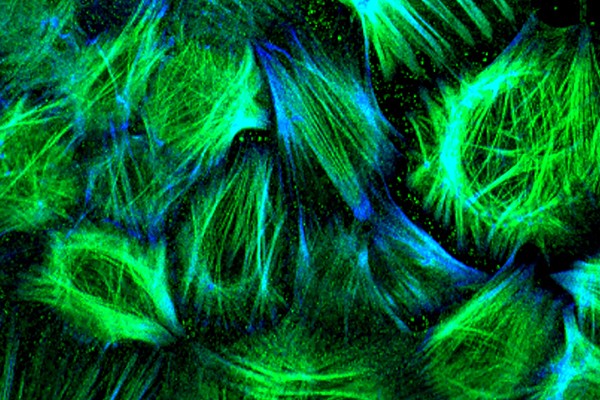 Image from Sue Menko, PhD, and Janice Walker, PhD: A 3D image of fibrotic cells associated with the lens fibrotic disease Posterior Capsule Opacification (PCO).
Connecting the Puzzle of Fibrotic Diseases
Up to 40 percent of disease mortality in developed countries results from fibrotic diseases, a family of conditions where the build-up of scar-like tissue interferes with the body’s normal function. These conditions can be systemic (such as systemic sclerosis and scleroderma) or affect individual organs such as the lung, liver, kidney, heart or eye. Because the mechanisms underlying these conditions are poorly understood and treatments are few, Jefferson has made a major commitment to basic and translational research on a broad range of fibrotic diseases.
Pulmonary Fibrosis (PF) causes progressive, potentially fatal scarring of the lung. PF results, in part, from the inability of aging lung epithelial cells to regenerate after injury. This may be because aging cells do not direct nutrients to areas at greatest need after injury. Ross Summer, MD, professor of medicine, has been working to determine the interconnections between cellular metabolism and injury repair. His studies on young cells’ metabolic adaptation to injury offer clues on how aging compromises the repair process—and suggests therapeutic approaches. For example, Dr. Summer has found that blocking lipid synthesis, alone, can cause fibrotic scarring in animal models; and, in counterpoint, has demonstrated that increased lipid production could reduce fibrosis-caused lung scarring significantly. He is now working to identify and test molecules that could have therapeutic effect by enhancing or protecting lipid synthesis.
Because scars that form in all injured tissues and organs are comprised primarily of collagen-rich fibrils, Andrzej Fertala, PhD, professor of orthopaedic surgery, has been developing an antibody to block an early step in fibril formation. Having shown the antibody to be effective at blocking fibril production in a mouse model of pulmonary fibrosis and a rabbit model of arthrofibrosis, Dr. Fertala is now fine-tuning it to create a therapeutic-grade biologic. Teaming with orthopaedic surgeons, Dr. Fertala’s group is also testing the possibility of employing the anti-fibrotic antibody to block fibrosis of injured peripheral nerves, which hampers the nerve regeneration process.
Cataract surgery often results in a fibrotic disease known as Posterior Capsule Opacification (PCO), which can lead to loss of vision. Sue Menko, PhD, professor of pathology, anatomy and cell biology, developed a chick embryo model of PCO and uses it to identify cell-signaling pathways involved in fibrosis and to study other mechanisms of wound healing and fibrosis. These studies have led to a series of significant discoveries. For example, she and Janice Walker, PhD, assistant professor of pathology, anatomy and cell biology, found that the lens harbors a resident population of vimentin-rich mesenchymal cells that migrate to the wound edge where they play a role in wound repair. Spurred by their encounter with a microenvironment of increased rigidly, these cells differentiate to myofibroblasts—the principal cell type associated with fibrosis. Subsequently, the researchers found that injury spurs the release of the protein vimentin, which binds to mesenchymal cells at the wound and helps signal the shift into a myofibroblast and the development of fibrosis. These findings may provide novel targets for development of new treatments for PCO.
Until very recently, the lens had been considered not susceptible to the immune processes normally associated with wound repair; most investigators assumed that immune cells played no role in PCO. However, using a lens-specific knockout mouse model, Dr. Menko’s team found that degeneration of the lens activates an immune response both to the lens and throughout the eye (including cornea, vitreous humor and retina) that contributes to development of fibrosis associated with cataractogenesis. Additionally, they demonstrated that the lens is indirectly connected to the lymphatic system, providing a potential mechanism for the immune surveillance. Their findings have broad implications for understanding disease and injury-repair processes throughout the eye.
Children with a grave skin disorder known as butterfly disease (or epidermolysis bullosa) develop severe and chronic blisters and fibrosis within their connective tissues. The condition leads to club-like appendages where the skin grows over the fingers or toes. Andrew South, PhD, associate professor of dermatology and cutaneous biology, has shown that fibrosis also leads to an aggressive, often fatal, form of skin cancer in butterfly children. Recently, Dr. South and colleagues pinpointed how fibrosis develops in butterfly children—and demonstrated that a specific molecule interferes with the process and reduces fibrosis in a tissue-engineered model. Now his team is working to develop the molecule as a potential therapy for the debilitating condition.
A Distinguished Career Honored—and the Work Continues
 Jouni Uitto, PhD Professor and Chair of Dermatology and Cutaneous Biology
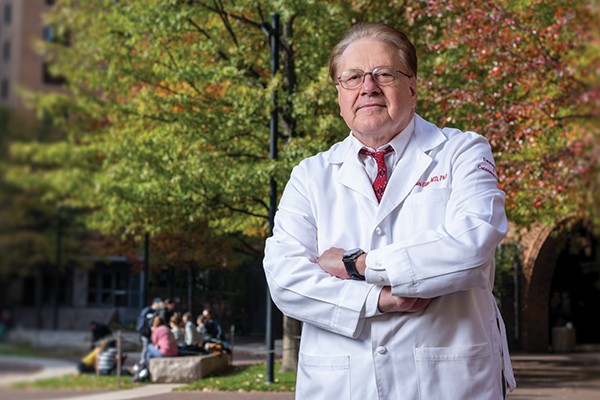
In 2019, Dr. Jouni Uitto, professor and chair of Dermatology and Cutaneous Biology, received the World Congress of Dermatology’s Alfred Marchionini Medal in Gold. The Medal is bestowed every four years and is the most prestigious award in international dermatology. The award is a recognition of Dr. Uitto’s long and distinguished career, in which he has published nearly 900 peer-reviewed articles, textbook chapters and reviews—including 30 new publications over the past year.
Dr. Uitto is particularly renowned for his research on a group of genetic diseases called epidermolysis bullosa (EB), which manifest at birth with extensive skin blistering and scarring. In its most severe form, EB can be fatal during a child’s first weeks or months of life. His lab discovered the disease’s genetic basis: mutations in the collagen7A1 gene undercut the production of a protein necessary for keeping layers of the skin together. The lab then developed a transgenic mouse model of EB, which became a platform for development of treatment approaches. Today, five gene therapy approaches in clinical trial have their origins in Dr. Uitto’s work.
He is also driving advances on the rare childhood disease pseudoxanthoma elasticum (PXE)—where mineral build-up in elastic fibers of the skin and blood vessels lead to formation of plaques in blood vessels and eyes that cause heart attacks and blindness. Collaborating closely with parents of children with PXE, Dr. Uitto gathered DNA samples that enabled his lab to identify the responsible genes. Today, clinicians can make a prognosis based on a patient’s specific set of mutations, and research continues to develop and test effective therapies.