Identifying Biomarkers to Assess Pediatric Spinal Injury
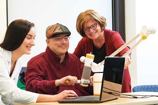 MJ Mulcahey, PhD, met Miki Greenstein, pictured, soon after he had a spinal cord injury at the age of 13. She has worked with him for 20 years now, and he is a frequent participant in her research studies. Here, she and student Samantha Burke work with Miki on a SaeboReJoyce rehabilitation workstation that helps with arm and hand training.
"This work could fill a major gap in pediatric care."
- Mary Jane Mulcahey, PhD
Professor of Occupational Therapy
Diagnosing and assessing spinal cord injury (SCI) in children and adolescents has long been a challenging task, as has been the application of relevant and established outcomes measures for these patients. “In adults, the neurological consequence of SCI is determined by physically examining 56 sites on the patient’s body—examinations that require full participation and cognitive awareness,” explains Mary Jane Mulcahey, PhD, Professor of Occupational Therapy.
“But we showed in a study of 236 children with SCI that children younger than 6 years—and some up to 10 years old—are not able to participate effectively. The clinical assessment of SCI is also challenging with adults who have brain injury or are in a coma or medical-induced sedation. We need to develop imaging biomarkers that tell us—regardless of a patient’s age or mental capacity—the precise extent of damage.”
With a team of biostatisticians, engineers, neuro-radiologists, neuroscientists, occupational therapists and physicists, Dr. Mulcahey has been developing and validating spinal cord imaging biomarkers and testing the feasibility of neuroimaging to predict SCI outcomes. Their proof-of-concept study employed diffusion tensor imaging (DTI) to effectively analyze both direct and indirect spinal cord damage in children and youth between birth and 21 years old at the time of injury.
Since then, funded by the NIH, the team has successfully employed an expanding array of imaging technologies to visualize in detail the original injury and subsequent pathologies. Those techniques have included spinal cord cross sectional area, which offers a structural biomarker for spinal cord atrophy and related pathology; and fiber tractography, which provides a 3D reconstruction of neural tracts using data collected by DTI.
Throughout the studies, the researchers have correlated biomarker-focused discoveries and observations of patients’ clinical presentation. Now they are ready to take the important next step: developing and testing projections for individual patients, based on comprehensive imaging studies of newly injured patients and multi-year tracking of their progress.
“To our knowledge, we are the only group seeking to define and validate pediatric SCI imaging biomarkers,” Dr. Mulcahey observes. “We are excited about this work because it could fill a major gap in pediatric care—and, potentially, inform better treatment of adult SCI too.”